Home
/ Plant Cell Organelles Under Microscope / Plant Cell Structure Read Biology Ck 12 Foundation : However, hooke observed dead cells under the microscope as cork is made up of dead cells.
Plant Cell Organelles Under Microscope / Plant Cell Structure Read Biology Ck 12 Foundation : However, hooke observed dead cells under the microscope as cork is made up of dead cells.
Plant Cell Organelles Under Microscope / Plant Cell Structure Read Biology Ck 12 Foundation : However, hooke observed dead cells under the microscope as cork is made up of dead cells.. Ribosomes, endoplasmic reticulum, lysosomes, centrioles, and golgi bodies due their smaller size they were discovered after the introduction of electron microscope. They are responsible for photosynthesis , for storage of products such as starch, and for the. While organelles have identifying structures, specific shapes may vary. For organelles that can be seen under the light microscope are mainly the protoplasm: Nucleus, cytoplasm, cell membrane, chloroplasts and cell wall (last 2 organelles are only present in plant cells).
The cells may conatin the following cell organelles depending upon wether it is a plant or animal cell We say cells are microscopic because they can only be seen under a microscope. The organelles in a cheek cell that are not visible under a light microscope are the ribosomes. The basic units of life. They are responsible for photosynthesis , for storage of products such as starch, and for the.
Cell Structure A Level The Science Hive from images.squarespace-cdn.com Chloroplasts berdurasi 18.000 detik pada 25fps. The organelles in a cheek cell that are not visible under a light microscope are the ribosomes. We say cells are microscopic because they can only be seen under a microscope. Of course, these epithelial cells in your mouth can be observed under a microscope in its high power. Are you looking for information on plant cell organelles and their functions? Animal cells and plant cells. Which of the following cell structures can you see under a light microscope? Nucleus, cytoplasm, cell membrane, chloroplasts and cell wall (last 2 organelles are only present in plant cells).
The vacuoles contain cell sap, which is a solution of sugars, amino acids, mineral salts, waste chemical and anthocyanin pigments.
Under and electron microscope a lattice like structure is visible. Every cell in your body contains organelles (structures that have specific functions). Even though plants and animals belong to eukaryotes. These organelles use specialized microtubules called spindle fibers to pull one copy of each condensed chromosome to either side of the cell. Of course, these epithelial cells in your mouth can be observed under a microscope in its high power. However, hooke observed dead cells under the microscope as cork is made up of dead cells. Learn how to make an animal cell cake! Just like organs in the body, each organelle contributes in its in fact, without microscopes, we wouldn't even know that organelles existed! Consists of a thin layer of amphipathic lipids which spontaneously arrange so more hydrophilic regions associate with it's main functions are storage, energy, and manufacturing. Chloroplasts berdurasi 18.000 detik pada 25fps. Are similar in that they are both eukaryotic cells and have similar organelles. Cell wall (in plants, not animals), ribosomes, plasma membrane, flagella, chromosomes, cytoplasm(made of cytosol, a semifluid substance). Plant cells are eukaryotic cells with a true nucleus along with specialized structures called organelles that carry out certain specific functions. some of these differences can be clearly understood when the cells are examined under an electron microscope.
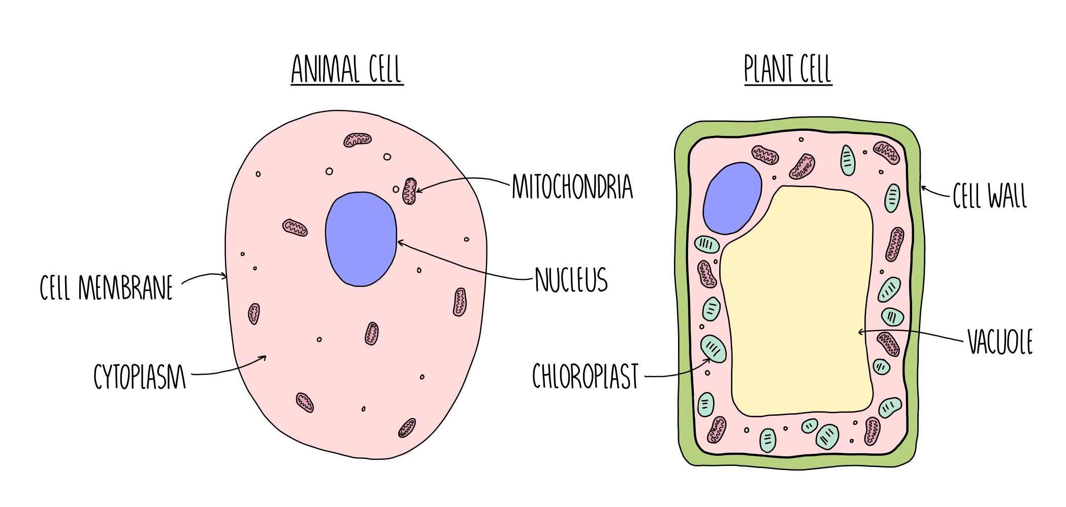
Major differences between a plant cell and on animal cell are (i) presence of chloroplast in plant name the two organelles in a plant cell that contain their own genetic material and ribosome. Are similar in that they are both eukaryotic cells and have similar organelles. Animal cells • there are a number of differences between plant and animal cells when they are viewed under a microscope • cell size and shape of animal and plant cells differ • some organelles are found only in one cell type, but not in both (cell wall and chloroplast in plant cells. Under and electron microscope a lattice like structure is visible. Every cell in your body contains organelles (structures that have specific functions).
What Are The Visible Plant Animal Cell Organs On Light Microscope Quora from qph.fs.quoracdn.net Video 4k dan hd siap untuk segala nle. Nucleus, cytoplasm, cell membrane, chloroplasts and cell wall (last 2 organelles are only present in plant cells). Consists of a thin layer of amphipathic lipids which spontaneously arrange so more hydrophilic regions associate with it's main functions are storage, energy, and manufacturing. Cells consist of cytoplasm enclosed within a membrane, which contains many biomolecules such as proteins and nucleic acids.2 most plant and animal cells are only visible under a light microscope, with dimensions between 1 and 100 micrometres.3 electron microscopy gives a much higher. They are responsible for photosynthesis , for storage of products such as starch, and for the. Animal cells • there are a number of differences between plant and animal cells when they are viewed under a microscope • cell size and shape of animal and plant cells differ • some organelles are found only in one cell type, but not in both (cell wall and chloroplast in plant cells. When you look at a cell in telophase under a microscope, you will see the dna at either pole. If a plant cell has chloroplasts and starch grains as seen under microscope it means the cell has synthesized its own food by photosynthesis.
However, most organelles are not clearly visible by light microscopy, and those.
However, most organelles are not clearly visible by light microscopy, and those. If a plant cell has chloroplasts and starch grains as seen under microscope it means the cell has synthesized its own food by photosynthesis. These organelles are responsible for protein synthesis. Appearance —under a microscope, normal cells and cancer cells may look quite different. Are you looking for information on plant cell organelles and their functions? Hence, cells exhibit cell division. The organelles in a plant cell vary in size. Some organelles are visible with a compound light microscope, while other organelles can be seen only plant cell organelles that are invisible under a compound light microscope include mitochondria, ribosomes, endoplasmic reticula, and golgi bodies. Resolving power is the ability to distinguish between separate. The basic units of life. A cell is a very tiny structure which exists in living bodies. Organelles can be divided into three types. A micrograph is a photo or digital image taken through a microscope to show a magnified image of a specimen.
In plant cells vacuoles are large, bounded by a single unit membrane called tonoplast. They have green pigment called chlorophyll which allow them to use the energy of. These organelles use specialized microtubules called spindle fibers to pull one copy of each condensed chromosome to either side of the cell. Ribosomes, endoplasmic reticulum, lysosomes, centrioles, and golgi bodies due their smaller size they were discovered after the introduction of electron microscope. It also has a very high resolving power.
Cell And Organelles Dr Jastrow S Electron Microscopic Atlas from www.drjastrow.de The short answer is it depends on the microscope. A mitochondrion surrounded by rough er under a transmission electron microscope. These organelles use specialized microtubules called spindle fibers to pull one copy of each condensed chromosome to either side of the cell. Animal cells • there are a number of differences between plant and animal cells when they are viewed under a microscope • cell size and shape of animal and plant cells differ • some organelles are found only in one cell type, but not in both (cell wall and chloroplast in plant cells. Hence, cells exhibit cell division. It may still be in its condensed state or thinning out. However, most organelles are not clearly visible by light microscopy, and those. Pilih dari berbagai adegan serupa.
The short answer is it depends on the microscope.
However, hooke observed dead cells under the microscope as cork is made up of dead cells. A mitochondrion surrounded by rough er under a transmission electron microscope. A mother cell divides to produce daughter cells. These organelles use specialized microtubules called spindle fibers to pull one copy of each condensed chromosome to either side of the cell. A micrograph is a photo or digital image taken through a microscope to show a magnified image of a specimen. Most organelles are common to both animal and plant cells. It may still be in its condensed state or thinning out. Nucleus, cytoplasm, cell membrane, chloroplasts and cell wall (last 2 organelles are only present in plant cells). A cell is a very tiny structure which exists in living bodies. Organelles can be divided into three types. Major differences between a plant cell and on animal cell are (i) presence of chloroplast in plant name the two organelles in a plant cell that contain their own genetic material and ribosome. Cells and organelles worksheet inspirational cell organelle matching game printable | chessmuseum template library. Chloroplasts (green plastids) are amazing organelles found in the cells of green plants and algae.
Dapatkan video stok chloroplast under a microscope plant cell organelles. Cells and organelles worksheet inspirational cell organelle matching game printable | chessmuseum template library.
Share :
Post a Comment
for "Plant Cell Organelles Under Microscope / Plant Cell Structure Read Biology Ck 12 Foundation : However, hooke observed dead cells under the microscope as cork is made up of dead cells."


Post a Comment for "Plant Cell Organelles Under Microscope / Plant Cell Structure Read Biology Ck 12 Foundation : However, hooke observed dead cells under the microscope as cork is made up of dead cells."